Se habla español | We treat emergencies
1250 W. Lake St, Unit #20, Addison, IL 60101Se habla español | We treat emergencies
1250 W. Lake St, Unit #20, Addison, IL 60101
An intraoral camera is a compact, pen-like imaging device designed to capture high-resolution, full-color images of the teeth and surrounding soft tissues. By bringing the camera into the mouth, clinicians can illuminate and magnify areas that are difficult to see with the naked eye. The result is a crisp visual record that highlights surface anatomy, early wear patterns, cracks, discoloration, and subtle gum changes before they become more serious problems.
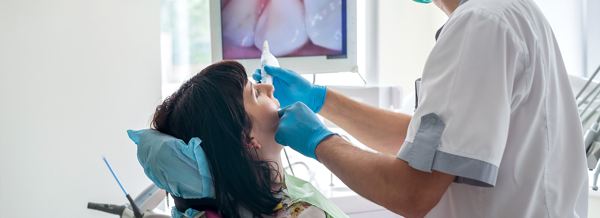
Unlike traditional film or standard digital radiographs, intraoral camera images show surface details in real time and in color, which makes them especially useful for assessing enamel defects, restorations, and the condition of soft tissues. Images can be displayed on a chairside monitor so patients and clinicians see the same view simultaneously. This shared display helps demystify clinical findings and supports clearer discussions about next steps in care.
Because the device is noninvasive and easy to maneuver, intraoral cameras have become a routine part of many preventive and diagnostic appointments. The images they produce can be saved to the patient’s digital chart, providing a historical record that helps track changes over time and supports continuity of care across visits.
One of the most immediate benefits of intraoral cameras is their ability to improve communication. When clinicians can show patients an up-close view of a specific tooth or gum area, explanations become concrete rather than abstract. Patients often leave appointments with a clearer understanding of their oral health because they can see what the dentist sees, rather than relying solely on verbal descriptions.
This visual approach encourages informed decision-making. When treatment recommendations are accompanied by images, patients are better equipped to weigh options and understand the urgency or timing of care. For many people, seeing an image makes it easier to prioritize preventive measures and accept restorative work when indicated.
From an educational standpoint, intraoral camera images help reinforce good home-care habits. Practitioners can point out plaque accumulation sites, areas prone to brushing abrasion, or the effects of habits such as grinding. When patients see these issues visually, they are often more motivated to adopt targeted hygiene strategies.
In clinical practice, intraoral cameras are versatile tools that support a wide range of procedures. They help identify early-stage caries, evaluate margins of existing restorations, and detect hairline fractures that might not show on X-rays. The ability to capture detailed surface images complements radiographic data, giving clinicians a more complete picture of oral health.
The cameras are also valuable in periodontal assessments, enabling careful inspection of gingival tissues for signs of inflammation, recession, or unusual lesions. For prosthetic and cosmetic planning, photographic documentation can guide shade selection, margin design, and communication with dental laboratories to ensure predictable results.
When multiple providers are involved in a patient’s care, intraoral images facilitate clearer referrals and treatment coordination. Images saved to the chart can be shared with specialists or a laboratory to communicate exact findings and expectations, reducing ambiguity and improving clinical outcomes.
Modern dental practices increasingly rely on integrated digital systems, and intraoral camera images are a key component of that workflow. Images captured during an exam are typically stored in the patient’s electronic health record alongside radiographs and clinical notes. This centralization makes it easy to review previous images, compare changes, and document the progression or resolution of conditions.
Secure image storage also supports continuity of care across providers. When a restorative case requires a laboratory or a specialist consultation, clinicians can export or securely transmit images to provide a clear visual baseline. That precision reduces the risk of miscommunication and supports more predictable restorative or surgical planning.
For practices that maintain a patient portal or provide educational resources, selected images can be used to illustrate home-care instructions or follow-up needs. Because these photos are objective and time-stamped, they serve as reliable documentation for clinical decisions while keeping the focus on evidence-based care.
From a quality assurance perspective, intraoral photography supports clinical audits and case review. Clinicians can review images to verify treatment outcomes, refine diagnostic approaches, and track long-term results, all of which contribute to higher standards of care.
An intraoral camera exam is quick and comfortable. During a routine checkup or a focused diagnostic visit, the clinician or hygienist will gently move the camera around the mouth while watching the live feed on a monitor. Because the device is small and noninvasive, most patients experience little to no discomfort—just a clearer view of their own teeth and gums.
Images taken during the exam are reviewed with the patient in real time. The clinician will point out areas of interest, explain their observations, and discuss how the findings relate to overall oral health. Patients often find this collaborative review helpful, as it allows time to ask questions and consider personalized care options based on visible evidence.
After the exam, key images are saved to the patient’s chart for future reference. These photos provide a visual baseline that can be revisited at subsequent visits to assess whether recommended treatments or home-care adjustments are producing the expected improvements. In this way, intraoral cameras support both immediate diagnostics and long-term monitoring.
If additional treatment is recommended, the clinician will explain the rationale using the images as a guide. Whether the next step is a simple preventive measure, a restorative procedure, or a specialist referral, the photographs help ensure that patients understand the reasoning behind clinical recommendations.
At Addison Dental Studio, we view intraoral imaging as an essential part of modern, transparent dental care. By combining high-quality visuals with clear explanations, we help patients make informed choices and maintain healthier smiles. If you have questions about intraoral cameras or how they might be used during your visit, please contact us for more information.
An intraoral camera is a small, pen-shaped device that captures high-resolution, full-color images of teeth and soft tissues inside the mouth. It allows clinicians to magnify and illuminate areas that are difficult to see with the naked eye, producing clear visual records of surface anatomy, cracks, wear patterns, and discoloration. These images give a more complete view of oral health and frequently reveal early signs of problems before they progress.
Because images are captured in real time and displayed on a monitor, intraoral cameras improve diagnostic accuracy and support patient education. The visual evidence helps practitioners explain findings clearly and document conditions for future comparison. Over time, saved images create a chronological record that aids in monitoring changes and planning appropriate care.
Intraoral cameras capture detailed color images of surface anatomy, while dental X-rays reveal internal structures such as tooth roots and bone. The two tools are complementary: camera images show enamel defects, restoration margins, and soft tissue issues in color and high magnification, whereas radiographs provide information about underlying decay, bone level, and hidden pathology. Using both modalities gives clinicians a fuller diagnostic picture.
An intraoral camera is nonradiographic and provides immediate visual feedback without exposure to X-rays, making it especially useful for explaining findings to patients. Radiographs remain essential for detecting problems that are not visible externally, so clinicians typically use cameras alongside X-rays rather than as a replacement. Together they enhance diagnostic confidence and treatment planning.
Intraoral cameras are effective at identifying early-stage enamel defects, cracks, staining, and deterioration at the margins of existing restorations. They excel at revealing surface irregularities and subtle changes in soft tissues, such as inflammation, recession, or unusual lesions that warrant further evaluation. Because of their magnification, cameras can also help locate plaque accumulation and areas prone to abrasion.
The camera is particularly useful for monitoring fractures or small defects that may not immediately appear on radiographs, and for documenting the condition of crowns, bridges, and veneers. When images indicate concerning findings, clinicians can recommend targeted diagnostic steps or refer the patient for additional testing to confirm the diagnosis. Clear photographic documentation supports more precise clinical decision-making.
An intraoral camera exam is quick, noninvasive, and generally comfortable for most patients because the device is small and designed for intraoral maneuvering. The camera is simply moved around the mouth while the clinician or hygienist watches a live feed, and most people experience little to no discomfort during the procedure. There is no radiation involved with camera imaging, which makes it a safe adjunct to other diagnostic tools.
Clinicians follow standard infection control protocols when using intraoral cameras, including barrier protection and cleaning procedures between patients. If a patient has a strong gag reflex or other sensitivity, the clinician can adjust technique or pacing to maintain comfort. The overall safety profile and ease of use make the camera a routine part of many preventive and diagnostic visits.
When clinicians display intraoral images on a chairside monitor, patients see the exact view the dentist is using to make clinical observations. This shared visual reference turns abstract explanations into concrete examples, helping patients understand the nature and urgency of findings. Visual documentation enables more informed conversations about treatment options and home-care recommendations.
Images can also be annotated or revisited during follow-up visits, reinforcing key teaching points and motivating patients to adopt targeted hygiene strategies. By making dental conditions visible and time-stamped, photography supports transparent care discussions and strengthens the partnership between patient and clinician at Addison Dental Studio.
Yes. Intraoral images provide detailed documentation that informs restorative margin design, shade selection, and cosmetic planning by revealing fine surface details and tissue relationships. Photographs of existing restorations and adjacent structures help clinicians identify where adjustments or replacements are needed and guide communication with dental laboratories. This level of detail supports more predictable aesthetic and functional outcomes.
For complex restorative or prosthetic cases, sequential images allow the team to track progress from initial presentation through provisional and final stages. The photographic record helps ensure that laboratory technicians and specialists receive accurate visual information, which reduces ambiguity and contributes to treatment precision.
Intraoral images are typically saved directly to the patient’s electronic health record alongside radiographs and clinical notes, creating a centralized and time-stamped visual history. This digital storage makes it simple to retrieve past images, compare changes over time, and document the effectiveness of treatments or home-care interventions. Centralized records also improve continuity of care when multiple providers are involved.
Secure storage practices and access controls are used to protect patient privacy while allowing authorized clinicians to review images as needed. When images are needed for case planning or specialist consultation, they can be exported or securely transmitted with appropriate safeguards in place. Maintaining an organized photographic archive supports long-term clinical oversight and quality assurance.
Yes, intraoral images are often shared with specialists or dental laboratories to improve communication and treatment coordination when a referral or laboratory fabrication is required. Photographs provide objective visual context that complements written notes and radiographs, allowing receiving clinicians or technicians to understand specific findings and desired outcomes. Clear imagery reduces the risk of miscommunication and helps align expectations across providers.
Clinicians follow privacy and security protocols when transmitting images, ensuring that patient information is shared only with authorized parties and for legitimate clinical reasons. Sharing high-quality images supports more efficient collaboration and contributes to predictable restorative and surgical planning.
No special preparation is typically required for an intraoral camera exam beyond normal oral hygiene. It can be helpful to arrive with clean teeth so that plaque and surface debris do not obscure diagnostic images, but clinicians can capture usable photographs even when some buildup is present. If you have removable appliances, you may be asked to remove them briefly to allow a complete view of the tissues.
If you have concerns about gag sensitivity or strong oral reflexes, let the dental team know when scheduling or at check-in so they can adjust technique and timing to keep you comfortable. The clinician will explain the process at the start of the visit and review images with you in real time to answer any questions that arise.
Addison Dental Studio uses intraoral imaging as part of a systematic approach to document baseline conditions and track changes during and after treatment. Sequential images are compared over time to evaluate healing, the stability of restorations, and the effectiveness of hygiene or preventive measures. This photographic evidence enhances clinical audits and supports evidence-based adjustments to care plans.
By integrating images into the electronic chart, clinicians ensure that follow-up decisions are informed by objective, time-stamped visuals. Patients can review their own progress during visits, which helps reinforce adherence to recommended care and clarifies next steps when additional treatment or maintenance is indicated.
Quick Links
Contact Us