Se habla español | We treat emergencies
1250 W. Lake St, Unit #20, Addison, IL 60101Se habla español | We treat emergencies
1250 W. Lake St, Unit #20, Addison, IL 60101
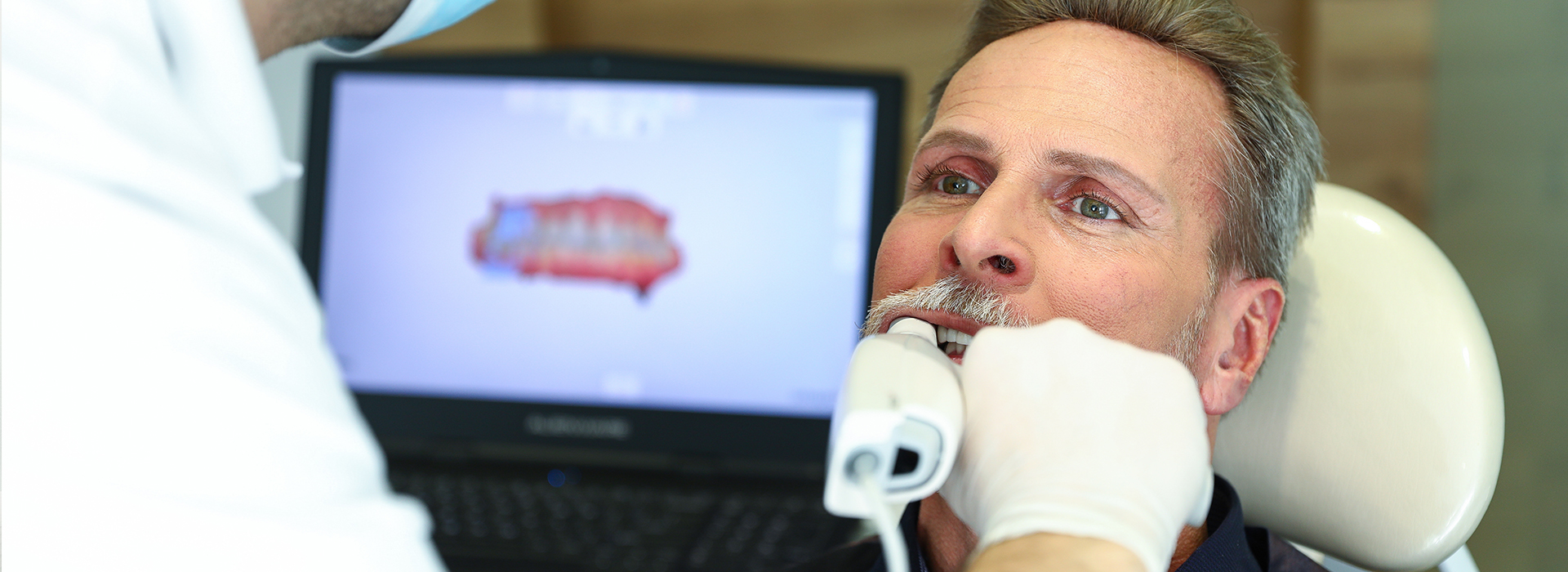
Digital impressions replace traditional putty-and-tray impressions with high-resolution optical scans captured inside the mouth. A handheld intraoral scanner collects thousands of tiny images or laser measurements in seconds and assembles them into a precise three-dimensional model of the teeth and surrounding soft tissues. That model can be viewed immediately on a monitor, rotated, and examined from multiple angles to evaluate margins, contacts, and occlusion before any restorative work begins.
The scanner records surface detail in micron-level increments, producing a file format that is compatible with modern dental laboratory systems and computer-aided design/manufacturing (CAD/CAM) tools. Because the data are digital from the start, they can be trimmed, annotated, or combined with other diagnostic data—like bite registrations or intraoral photos—without losing fidelity. This digital-first workflow reduces steps that traditionally introduced variation and error.
From a patient perspective, the process feels more clinical and less intrusive than conventional impressions. For clinicians, it provides instant feedback: areas that need rescanning are obvious on the screen, eliminating multiple physical retakes later. The result is a streamlined, repeatable method for capturing an accurate representation of a patient’s mouth that integrates cleanly with modern restorative and orthodontic workflows.
Many patients report greater comfort with digital scanning because there are no bulky impression trays or viscous materials sitting in the mouth. People with strong gag reflexes, anxiety about dental procedures, or difficulty keeping still benefit especially from the quick, non-invasive scanning approach. Because scans typically take only a few minutes per arch, appointments are shorter and less stressful for both patient and clinician.
Digital impressions also improve predictability of treatment. Clinicians can immediately assess whether the scan captures adequate detail around crown margins, undercuts, or adjacent restorations. When something needs correction, rescanning a small area takes seconds rather than having to repeat an entire traditional impression. That real-time assurance reduces the chance of surprises when the restoration is tried in.
For patients receiving crowns, bridges, or clear aligners, predictability translates into fewer adjustments and quicker final outcomes. The improved fit and better initial adaptation of restorations mean fewer follow-up visits for remakes or extensive chairside adjustments, which enhances overall satisfaction without introducing subjective claims about outcomes.
One of the biggest operational advantages of digital impressions is the speed of communication with dental laboratories. Digital files can be uploaded or transmitted securely to a preferred lab in minutes, removing mail time and the uncertainty of physical impressions traveling between offices. Labs can receive, evaluate, and start planning restorations almost immediately after the scan is approved.
Because files are standardized, dental technicians can work directly in CAD/CAM software to design restorations, plan implant prosthetics, or prepare models for 3D printing. This reduces the number of manual steps involved in analog model fabrication and allows clinicians to collaborate closely with technicians on adjustments before any physical piece is fabricated. The smoother handoff typically reduces turnaround variability and supports more predictable scheduling.
Digital workflows also facilitate better documentation. Every scan is archived as a digital record that can be retrieved for future treatment planning, comparison over time, or forensic review if needed. The ability to keep an accurate, time-stamped database of oral scans enhances continuity of care when multiple providers or specialists are involved.
Digital impressions are a cornerstone of chairside CAD/CAM systems that let clinics design and produce ceramic restorations in a single visit. After capturing a digital impression, a clinician or trained technician can design a crown, veneer, or inlay on a computer, then mill or 3D print the restoration in-house. For appropriate cases, this approach removes the need for temporary restorations and multiple appointments.
Not every situation is suitable for same-day fabrication, and clinical judgment remains essential when deciding whether to proceed chairside or involve a laboratory partner. When used properly, however, in-office restorations allow patients to leave with a definitive restoration sooner and with fewer appointments. That convenience is especially valuable for busy patients or when follow-up visits are difficult to schedule.
Even when restorations are fabricated by an outside lab, digital scanning supports rapid in-office adjustments and try-ins. The digital model can be used to preview occlusion and contacts before the physical restoration arrives, reducing the need for extensive chairside modification and ensuring the restoration integrates smoothly into the patient’s bite.
Numerous studies and clinical reports have shown that modern intraoral scanners produce levels of accuracy suitable for a wide range of restorative and orthodontic applications. For fixed prosthetics, the precise capture of preparation margins and adjacent structures helps technicians fabricate well-fitting restorations with predictable contacts and contours. For aligner therapy or implant planning, the combination of digital impressions with other digital imaging tools enhances overall treatment planning.
Beyond immediate restorative benefits, digital impressions create a durable digital archive of a patient’s oral condition. These records are valuable for monitoring wear, tracking periodontal changes, evaluating the progression of conditions, and coordinating multidisciplinary care. Because the files are digital, they are easy to share securely with specialists or to import into practice management and imaging systems.
Implementing digital impressions does require training, attention to scanning protocols, and equipment maintenance, but the clinical advantages—reduced remakes, clearer communication, and reliable records—make the investment worthwhile for many practices. When integrated thoughtfully into care pathways, digital impressions support higher standards of precision and consistency across a wide range of dental services.
Wrap-up: Digital impressions represent a modern, patient-friendly approach to capturing the shape and condition of teeth and supporting tissues. By combining comfort, clinical predictability, and streamlined laboratory communication, this technology helps practices deliver efficient, high-quality restorative and orthodontic care. If you’d like to learn more about how digital impressions are used in our office or whether they are right for your treatment, please contact us for more information.
Digital impressions replace traditional putty-and-tray techniques by using high-resolution optical scans captured inside the mouth. A handheld intraoral scanner collects thousands of images or laser measurements in seconds and assembles them into a precise three-dimensional model of the teeth and surrounding soft tissues. Clinicians can view and rotate that model immediately to evaluate margins, contacts and occlusion before any restorative work begins.
Scanners record surface detail in micron-level increments and produce file formats compatible with modern dental laboratory systems and CAD/CAM workflows. Because data are digital from the start, files can be trimmed, annotated or combined with bite registrations and intraoral photos without loss of fidelity. This digital-first workflow reduces analogue steps that historically introduced variation and error.
Traditional impressions rely on impression materials and physical models, which introduce handling, storage and shipping variables that can affect accuracy. Digital impressions eliminate many of those manual steps by creating a precise digital model at the point of care, reducing the risk of distortion and material-related error. The ability to inspect the scan instantly also lets clinicians identify and correct problems immediately.
Because rescans can target a small area rather than repeating an entire tray impression, digital workflows often reduce the number of physical retakes. Standardized digital files also simplify communication with dental laboratories and technicians, which can shorten turnaround variability. The net effect is a more controlled process from capture to fabrication.
Many patients find digital scanning less intrusive because it avoids large impression trays and viscous materials that can trigger gagging or discomfort. Scans typically take only a few minutes per arch, limiting the time a patient needs to remain still and reducing overall appointment length. The small, wand-like scanner is maneuvered gently around the teeth and soft tissues rather than filling the mouth with material.
Clinicians can show the scan on a monitor during or immediately after capture, which helps patients understand the treatment steps and what will be fabricated for their case. Quick rescans of localized areas remove the need for full repeat impressions and minimize additional chair time. For patients with a strong gag reflex or dental anxiety, the streamlined nature of scanning often makes the visit easier to tolerate.
Modern intraoral scanners capture surface anatomy and margin detail at micron-level resolution, enabling technicians to fabricate restorations with predictable contacts and contours. Accurate capture of preparation margins, adjacent teeth and occlusal relationships supports better-fitting crowns, bridges and implant restorations. When scans are combined with other digital records, such as CBCT or bite scans, planning for complex cases becomes more precise.
Immediate visual feedback during scanning lets clinicians address deficiencies on the spot, reducing the chance of remakes or extensive chairside adjustments later. Consistent scanning protocols and proper operator training are essential to realize these accuracy gains. When implemented correctly, digital impressions contribute to fewer surprises at try-in and more efficient restoration delivery.
Digital impressions streamline laboratory communication by producing files that can be transmitted electronically in minutes rather than relying on mail or courier services. Laboratories can receive, evaluate and begin designing restorations almost immediately after a scan is approved, eliminating transit delays and shrinking lead times. Digital files are standardized for CAD/CAM software, which reduces manual steps in model fabrication.
The digital handoff also enhances collaboration between clinicians and technicians because files can be annotated and adjusted virtually before any physical piece is produced. Archived scans serve as time-stamped records for future reference, simplifying case reorders or interdisciplinary planning. Overall, the workflow becomes more predictable and easier to schedule.
Digital impressions are a cornerstone of chairside CAD/CAM systems that allow clinics to design and produce ceramic restorations in a single visit for select cases. After capture, the clinician or a trained team member can design a crown, veneer or inlay on a computer and then mill or 3D print the restoration in-office. When clinical indications and material selection align, this approach can remove the need for temporary restorations.
Clinical judgment determines whether same-day fabrication is appropriate, since some cases require laboratory expertise or additional laboratory processes. Even when a restoration is sent to an external lab, a digital scan supports accurate previewing of occlusion and contacts and reduces the need for extensive chairside modification. Proper training and maintenance of in-office equipment are important to deliver reliable same-day outcomes.
Digital impressions are widely used for fixed prosthetics such as crowns, bridges, inlays and onlays, as well as for implant prosthetics and abutment design. Clear aligner therapy, digital removable appliance fabrication and occlusal splints also rely on accurate digital scans for predictable fit. The technology is increasingly used for diagnostic models, study planning and monitoring changes over time.
Because the files integrate with CAD/CAM and 3D printing workflows, clinicians can use the same digital record for multiple purposes across restorative and orthodontic care. Digital impressions also facilitate interdisciplinary planning when specialists need to share precise anatomical data. Adoption continues to expand as software compatibility and scanner capability improve.
Digital scans are archived as electronic records that can be linked to a patient file and time-stamped for continuity of care. Secure transmission protocols, encrypted uploads and compliant file-handling practices are used to protect patient privacy when sharing scans with dental laboratories or specialists. Integrating scans into practice management and imaging systems reduces the need for duplicate records and supports coordinated treatment planning.
Practices typically implement data retention and backup strategies to ensure scans remain accessible for future treatment or comparison. Access controls and periodic audits help maintain the security of stored files. When transfers are necessary, industry-standard secure file transfer methods minimize the risk of unauthorized access.
During a digital scanning appointment, the clinician or trained assistant will explain the procedure and then use a small wand-like scanner to capture the teeth and surrounding tissues. Scanning each arch normally takes only a few minutes, and the clinician may ask the patient to reposition, bite down or move slightly to capture occlusion and bite registration. The process is noninvasive and generally requires minimal preparation from the patient.
After capture, the clinician reviews the digital model on a monitor and can perform quick rescans of any local areas that need more detail. The scan is then used to plan next steps, whether that means sending files to a laboratory or initiating a chairside CAD/CAM workflow. Patients often appreciate being able to see the model and better understand the proposed restorative steps.
At Addison Dental Studio, digital impressions are incorporated into restorative and orthodontic workflows to improve precision and coordination of care. The practice uses intraoral scanning to document preparation margins, occlusal relationships and soft tissue contours, which supports laboratory design and chairside fabrication when appropriate. Scans are integrated with other diagnostic records to form a complete digital treatment plan.
Using a digital-first approach allows the clinical team to identify areas that require refinement immediately and to collaborate efficiently with dental technicians or specialists. Archived scans provide a reliable record for follow-up care, monitoring changes over time and facilitating multidisciplinary cases. This systematic use of digital impressions supports consistent, predictable care pathways for patients.
Quick Links
Contact Us