Se habla español | We treat emergencies
1250 W. Lake St, Unit #20, Addison, IL 60101Se habla español | We treat emergencies
1250 W. Lake St, Unit #20, Addison, IL 60101
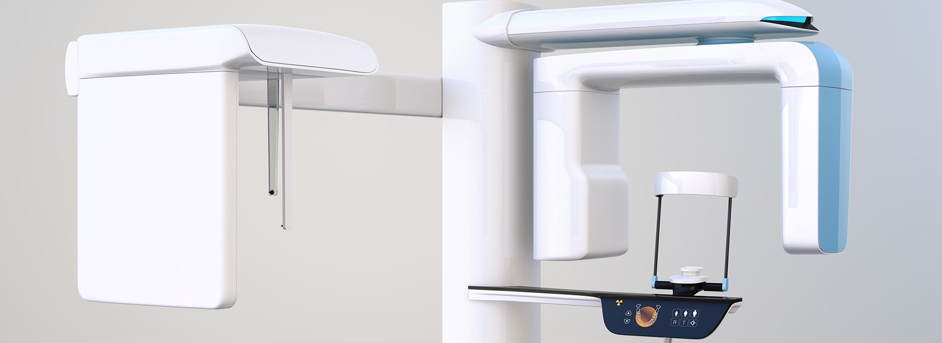
At Addison Dental Studio, we invest in diagnostic tools that make complex dental care more predictable, efficient, and comfortable. Cone-beam computed tomography (CBCT) is one of those technologies: a specialized 3D imaging system that gives clinicians an anatomically accurate view of teeth, bone, and surrounding facial structures without the distortion inherent to traditional two-dimensional X-rays.
CBCT scans are not used for every appointment, but when detailed spatial information is required—such as for implant placement, surgical planning, or evaluating complex root anatomy—they provide clarity that directly informs safer, more precise treatment. Our approach emphasizes both diagnostic value and patient safety, so each scan is ordered only when it will meaningfully improve care.
CBCT captures volumetric data that can be viewed slice by slice or reconstructed into three-dimensional models. This allows dentists to assess bone height and width, the location of nerves and sinuses, tooth root morphology, and pathological changes with a level of detail that plain films cannot match. These views help eliminate guesswork when anatomy is complex or when two-dimensional images are likely to overlap structures.
The technology provides selectable fields of view, so scans can be tailored to the diagnostic need—ranging from a focused scan of a single quadrant to a larger view of the entire jaw and midface. That flexibility reduces unnecessary exposure while delivering high-resolution detail in the region of interest.
Because CBCT data is spatially accurate, it supports measurements, surgical guides, and digital planning workflows. When planning an implant or evaluating a potential lesion, clinicians can measure bone dimensions and angulation directly from the scan, improving confidence when making clinical decisions.
One of the strongest advantages of CBCT is its role in preoperative planning. For dental implants, three-dimensional scans let clinicians visualize bone volume and density, avoid critical anatomical structures, and determine ideal implant size and angulation. When paired with digital surgical guides, CBCT-based plans can be translated into guided procedures that reduce chair time and lower the risk of surprises during surgery.
Beyond implants, CBCT assists in complex endodontic cases by revealing canal anatomy and identifying periapical pathology or root fractures that may be missed on standard X-rays. Oral surgeons use CBCT to map wisdom tooth relationships to the inferior alveolar nerve, enabling procedures to be carried out with greater precision and fewer complications.
CBCT also supports interdisciplinary care: orthodontists, periodontists, and otolaryngologists can use the same dataset to coordinate treatment. When multiple specialists collaborate using a shared, three-dimensional image, the result is a unified plan that prioritizes both function and long-term oral health.
A typical CBCT appointment is quick and straightforward. Patients are positioned either seated or standing depending on the machine; the scanner rotates around the head for a brief interval—usually under a minute—to capture the volumetric data. Because the scan is noninvasive and requires no contrast agents, most patients find the experience comfortable and unobtrusive compared with other imaging modalities.
Movement can degrade image quality, so technologists provide clear instructions to minimize motion and may use supports to stabilize the head. The short acquisition time reduces the likelihood of motion artifacts, which helps ensure the resulting dataset is diagnostically useful the first time.
Modern dental CBCT systems are designed to limit radiation exposure. By selecting the smallest field of view appropriate for the clinical question and using optimized exposure settings, clinicians adhere to the ALARA (As Low As Reasonably Achievable) principle while still obtaining the diagnostic information needed to plan care safely.
A CBCT dataset is only as valuable as the interpretation applied to it. Radiographic interpretation requires training and experience to distinguish normal variants from clinically significant findings. Our clinicians combine hands-on experience with advanced visualization tools that allow for multiplanar views, adjustable contrast, and measurement overlays to support accurate assessments.
When scans suggest findings beyond the scope of general dentistry—such as ambiguous lesions or complex airway concerns—we collaborate with specialists or oral and maxillofacial radiologists to ensure a thorough and responsible evaluation. That collaborative model helps ensure patients receive informed recommendations that reflect a comprehensive clinical perspective.
Software integration also enables digital workflows: 3D models derived from CBCT data can be merged with intraoral scans for restorative planning, prosthetic design, and the production of surgical guides. These digital tools streamline communication between clinician, laboratory, and patient, and they help translate diagnostic insight into precise clinical action.
Selecting a CBCT system is only the first step—ongoing maintenance, calibration, and staff training are essential to produce consistent, high-quality images. Our practice follows routine quality control procedures and invests in continuing education so every scan is captured and processed to meet current standards of care.
Clinical judgment guides the decision to image: we balance the benefit of additional diagnostic information against the imperative to minimize exposure. Imaging protocols are individualized based on the patient’s medical history, the specific diagnostic question, and the anatomical area under evaluation.
We also prioritize clear communication. Before ordering a scan, clinicians explain why the imaging is recommended, what it will reveal, and how the results will influence treatment. Patients are empowered to participate in decisions about their care with a clear understanding of the diagnostic value of CBCT scans.
In summary, CBCT is a powerful diagnostic tool that enhances precision, safety, and predictability in modern dental care. When used judiciously and interpreted by trained clinicians, three-dimensional imaging can directly improve planning for implants, surgical procedures, endodontic treatments, and other complex cases.
To learn whether CBCT is appropriate for your dental needs, please contact us for more information. Our team at Addison Dental Studio is available to answer questions and help determine the best diagnostic path for your treatment.
Cone-beam computed tomography (CBCT) is a specialized three-dimensional dental imaging technology that captures volumetric data of the teeth, jaws, and surrounding facial structures. Unlike traditional two-dimensional X-rays, CBCT records a cone-shaped beam as it rotates around the head, producing a dataset that can be reconstructed into multiplanar views and 3D models. This spatially accurate information helps clinicians visualize anatomy with greater detail for diagnosis and treatment planning.
CBCT images preserve relationships between structures, which enables precise measurements of bone dimensions, root morphology, and the location of critical anatomy such as nerves and sinuses. The technology offers selectable fields of view so clinicians can limit the scanned region to the area of interest. When used judiciously, CBCT provides diagnostic insights that plain films cannot supply while supporting more predictable clinical decisions.
Conventional dental X-rays produce two-dimensional projections that can superimpose structures and limit assessment of depth and spatial relationships. CBCT acquires volumetric data that can be sliced into axial, coronal, and sagittal planes or rendered as a three-dimensional model, eliminating much of the overlap seen on plain films. This difference makes CBCT particularly valuable when anatomy is complex or when precise measurements are required.
Another important distinction is field-of-view control: CBCT systems allow clinicians to select a focused scan area to reduce exposure and capture high-resolution detail where it is needed. While periapical and panoramic radiographs remain effective for many routine evaluations, CBCT is reserved for cases in which additional three-dimensional information directly affects treatment. The two modalities are complementary and are chosen based on the clinical question.
A CBCT scan is typically recommended when three-dimensional information will materially influence diagnosis or treatment planning, such as in implant placement, complex endodontic evaluation, surgical extractions, and assessment of jaw pathology. Clinicians also use CBCT for cases involving impacted teeth, atypical root anatomy, or suspected fractures that are not clearly visible on two-dimensional images. The decision to image is guided by clinical judgment and the principle of providing information that improves care.
CBCT may also be useful when interdisciplinary collaboration is required, for example when orthodontists, periodontists, or oral surgeons need a shared dataset to coordinate treatment. The scan protocol is individualized to the diagnostic need, selecting the smallest field of view and exposure settings that will yield adequate image quality. By tailoring scans to the question at hand, clinicians balance diagnostic benefit with patient safety.
For implant planning, CBCT offers precise visualization of bone volume, ridge morphology, and the relationship of proposed implant sites to vital anatomical structures such as the inferior alveolar nerve and maxillary sinuses. These measurements allow clinicians to determine implant length, diameter, and angulation with greater confidence than two-dimensional imaging permits. The three-dimensional view reduces the risk of surprises during surgery and supports selection of appropriate implant positions.
CBCT datasets can be integrated into digital planning software to create virtual implant placements and fabricate surgical guides that translate the plan into a guided procedure. This workflow enhances accuracy during osteotomy preparation and implant insertion, potentially shortening chair time and improving predictable outcomes. When combined with clinical assessment and prosthetic planning, CBCT-derived information contributes to a comprehensive, restorative-driven approach to implant care.
Modern dental CBCT systems are designed to minimize radiation exposure by offering selectable fields of view and adjustable exposure parameters that match the diagnostic need. Clinicians follow the ALARA principle—As Low As Reasonably Achievable—by selecting the smallest volume and lowest settings that will provide diagnostic quality images. When used appropriately, the radiation dose from a focused dental CBCT scan is generally modest compared with many medical CT exams.
Risk management also includes clinical justification and documentation: a scan is ordered only when the expected diagnostic benefit outweighs the exposure. Technologists use positioning aids and verbal instructions to limit motion artifacts and avoid repeat scans, and practices maintain routine quality assurance for equipment calibration. Patients with specific concerns about radiation should discuss them with the clinician so the imaging decision can be individualized.
A typical CBCT appointment is brief and noninvasive: the patient is positioned seated or standing depending on the unit, and the scanner rotates around the head for a short acquisition time, often less than one minute. No contrast agents or injections are required, and the procedure is generally well tolerated by most patients. Technologists provide instructions to remain still and may use head supports to reduce movement during the scan.
Because the acquisition is fast and automated, preparation is minimal; patients may be asked to remove metal accessories such as earrings, glasses, or removable prostheses that could create artifacts. After the scan, the dataset is processed for review, and clinicians will explain how the images will be used to inform diagnosis and treatment planning. Clear communication before imaging helps patients understand the purpose and expected benefits of the study.
Interpreting CBCT datasets requires training to distinguish normal anatomical variation from clinically significant findings and artifacts. In general practice, dentists with appropriate imaging training review the scans to address the referring question, using multiplanar views, adjustable contrast, and measurement tools to support assessment. For findings that extend beyond the scope of general dentistry—such as ambiguous lesions or complex airway concerns—collaboration with specialists or referral to an oral and maxillofacial radiologist is standard practice.
When interpretation is performed in-house, clinicians document relevant observations and explain their implications for treatment planning. Collaboration and second opinions are used when additional expertise is warranted, ensuring a responsible and comprehensive evaluation. These steps help translate imaging data into accurate diagnoses and informed recommendations for care.
CBCT data can be exported and combined with intraoral scans and digital prosthetic designs to create an integrated restorative and surgical plan. Software platforms allow clinicians to perform virtual implant placement, simulate outcomes, and design custom surgical guides that transfer the digital plan into the operatory. This integration improves communication among clinicians, laboratories, and patients by providing a shared visual reference for intended treatment.
The use of CBCT-derived guides does not replace clinical judgment but augments precision during implant osteotomies and complex surgeries. Digital workflows can reduce intraoperative guesswork and facilitate predictable prosthetic outcomes by aligning surgical placement with restorative goals. Proper case selection and validation of the guide in the clinic remain essential steps in this process.
While CBCT is a powerful diagnostic tool, it has limitations including susceptibility to metal artifacts, limited soft-tissue contrast compared with medical CT or MRI, and the potential for motion degradation if a patient cannot remain still. Some patients—such as pregnant individuals—require special consideration, and imaging should be deferred or justified carefully in these circumstances. The technology is not a universal replacement for all imaging modalities but rather a targeted tool for specific diagnostic questions.
Clinical judgment guides whether CBCT is the appropriate choice; factors such as the diagnostic question, patient cooperation, prior imaging, and alternative modalities all influence the decision. Imaging protocols are individualized to minimize unnecessary exposure while obtaining clinically useful information. When limitations are identified, clinicians may seek additional studies or specialist consultation to reach a definitive diagnosis.
At Addison Dental Studio, CBCT is applied selectively to cases where three-dimensional information will materially influence diagnosis and treatment planning, such as implant rehabilitation, complex endodontic assessment, and surgical guidance. The practice emphasizes equipment maintenance, staff training, and individualized imaging protocols so each scan yields diagnostically valuable data while adhering to radiation safety principles. Clear communication with patients about the rationale for imaging helps ensure informed decision-making.
Scans are interpreted by trained clinicians and, when appropriate, reviewed in collaboration with specialists to confirm findings and next steps. Integration of CBCT data with digital planning tools supports precise treatment workflows and coordinated interdisciplinary care. This measured approach positions the practice to deliver safer, more predictable outcomes rooted in accurate diagnostic information.
Quick Links
Contact Us